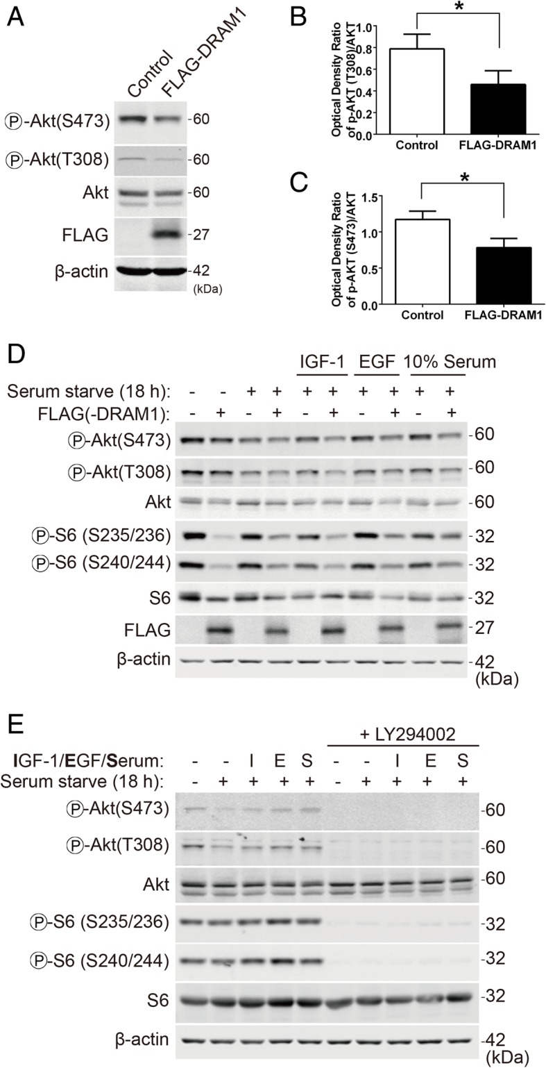
Fig. 3.

DRAM1 inhibits the activation of the PI3K-Akt pathway stimulated with growth factors and serum. a HEK293T cells were transfected with FLAG empty vector or FLAG-DRAM1 for 24 h and the protein levels of p-Akt (S473 and T308), FLAG and β-actin were detected with immunoblotting. b and c Quantitative analysis of the optical densities of p-Akt (T308, S473) in HEK293T cells transfected with FLAG-DRAM1. Data represent mean ± SEM of combined data from three independent experiments. d HEK293T cells were transfected with FLAG empty vector or FLAG-DRAM1 for 24 h, and then starved for another 18 h before stimulating with IGF-1 (5 ng/ml), EGF (50 ng/ml) and serum (10%) for 10 min. The protein levels of p-Akt (S473 and T308), Akt, p-rpS6 (S235/236, S240/244), rpS6, FLAG and β-actin were detected with immunoblotting. e HEK293T cells were transfected with FLAG empty vector or FLAG-DRAM1 for 24 h, and then starved for another 18 h before stimulated with IGF-1, EGF or serum (10%) for 10 min in the presence of or absence of LY294002 (50 μM). The protein levels of p-Akt (S473 and T308), Akt, p-rpS6 (S235/236 and S240/244), rpS6 and β-actin were detected by immunoblotting. *p < 0.05 vs control
