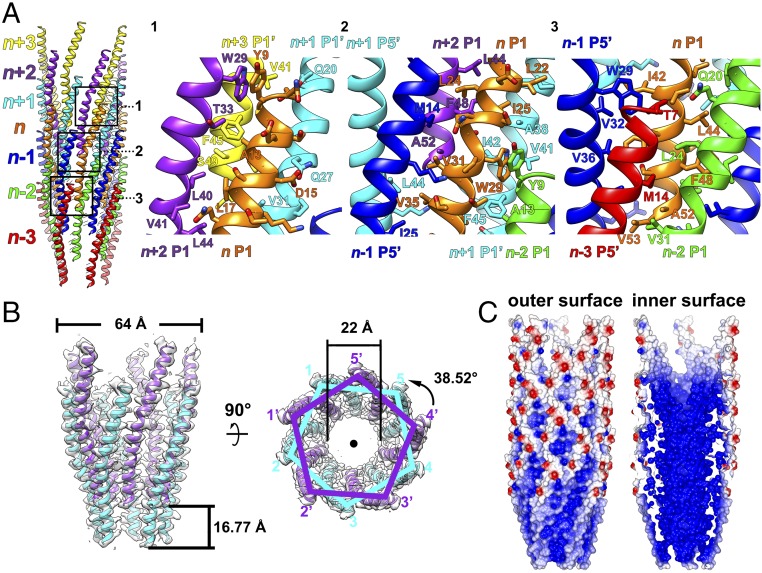
Fig. 2.
Helical assembly of the IKe capsid. (A, Left) Ribbon diagram showing an IKe capsid fragment of seven pentameric protein layers (n + 3, n + 2, n + 1, n, n − 1, n − 2, n − 3) in different colors. (A, Right) Ribbon and stick diagrams of numbered boxed areas in the Left panel showing the interactions of one p8 monomer (n P1, in orange) with neighboring subunits. The major coat proteins from the same pentameric protein layer are colored identically. (B) Ribbon and surface shadowed diagrams showing side (Left) and top (Right) views of two pentameric protein layers and their relationship via helical symmetry operators. (C) Surface electrostatic potential of the p8 helical shell. Negative and positive electrostatic potentials are colored red and blue, respectively.

