Fig. 5.
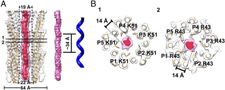
Structure of the inner ssDNA core. (A, Left) Ribbon and surface shadowed diagram showing the asymmetric reconstruction of the filamentous bacteriophage IKe calculated from the original raw images with the orientations determined for the inner DNA core. The helical shell is colored white, and the DNA core density contoured at a level of 17 σ is transparent and colored pink. The DNA core density contoured at a level of 26 σ is colored red. The model of the capsid is fitted in the map. (Middle and Right) Surface shadowed diagram showing the helical features of the inner DNA core and the left-handed helix. The left-handed helical pitch of the inner DNA core is ∼34 Å. (B) Ribbon and surface shadowed diagrams showing thin sections of the virus. The interactions between the capsid shell and the inner DNA core are mainly mediated through residues Lys51 and Arg43. The Arg43 residues are likely in contact with the DNA backbone. The densities from the shells are transparent and colored as described in A. The structure of IKe capsid shell is colored orange.

