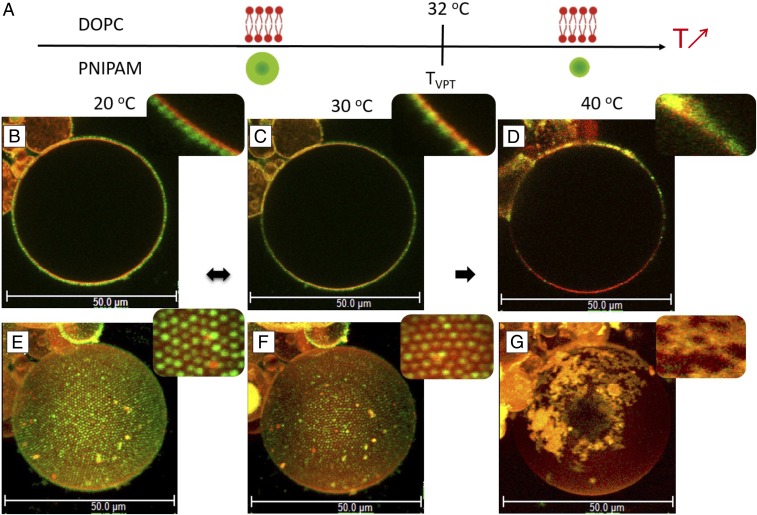
Fig. 2.
(A) Schematic representation of DOPC membrane and a thermoresponsive PNIPAM particle as a function of temperature. Below , microgel particles are swollen, soft, and hydrophilic, whereas above , they are collapsed, harder, and more hydrophobic. The lipid DOPC membrane is fluid over the whole temperature-interval investigated. (B–G) Fluorescence confocal micrographs showing a giant DOPC vesicle decorated with PNIPAM particles at 20 °C (B and E), 30 °C (C and F), and 40 °C (D and G). B–D, Insets reveal close-up views of lateral cross-sections showing the GUV–microgels contact line. (E–G) The 3D intensity projections of confocal -stacks [51.67 (x) 51.67 (y) 68.58 (z) ]. E–G, Insets show close-up views of particle organizations at the surface of the GUV.

