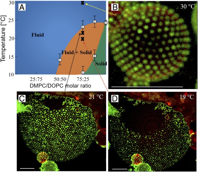
Fig. 4.
(A) Phase diagram of DOPC/DMPC lipids based on differential scanning calorimetry (DSC) data. (B) Fluorescence confocal micrographs of GUVs consisting of a mixture of DOPC/DMPC (75/25). At the highest temperature, 30 °C, when the whole bilayer forms a single fluid bilayer, the surface is homogeneously decorated with microgel particles. (C and D) When temperature is decreased, the lipid membrane separates into domains consisting of fluid and solid bilayer phases. The red fluorescent lipid analogue is only present in the fluid domains, and the solid domains appear black in the images. Images were taken at 21 °C (C) and 19 °C (D) showing the growth of the solid domains at lower temperature. It is clearly demonstrated that the microgel particles show strong preference of the fluid domains where it adsorbs to form 2D hexagonal crystals. Only very few particles were detected in the solid domains. (Scale bars: 10 m.)

