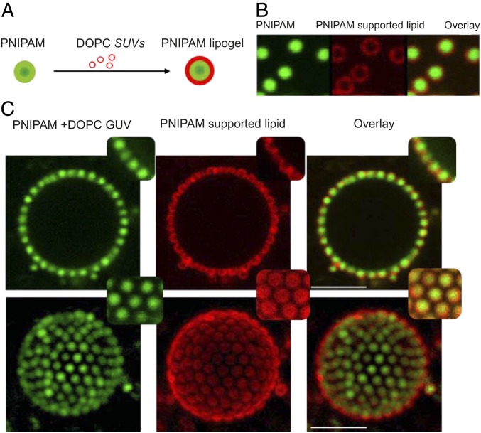
Fig. 5.
(A) Schematic illustration of lipogel preparation from PNIPAM particles and DOPC SUVs. (B) Fluorescence micrographs of lipid-rich lipogels adsorbed at a GUV, from left to right, showing green fluorescence of copolymerized FMA present in PNIPAM, red fluorescence of Rhod-PE present in DOPC lipid, and the overlay of the two channels. (C) The 2D and 3D micrographs of NBD-PE–labeled DOPC GUVs decorated with lipid-rich lipogels. From left to right: green fluorescence of copolymerized FMA in PNIPAM microgels and NBD-PE in GUVs, red fluorescence of Rhod-PE present in coated lipid, and the overlay of the two channels. Insets show close-up views of lateral cross-sections and at the top of the GUVs. Temperature: 20 °C. (Scale bars: 5 m.)

