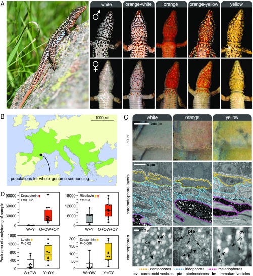
Fig. 1.
Color polymorphism in the European common wall lizard, P. muralis. (A, Left) Common wall lizard. (A, Right) Illustrations of the five discrete ventral morphs. These conspicuous colors likely function as visual ornaments implicated in sexual signaling. The yellow and orange colors are restricted to the ventral surface, and males and females exhibit marked differences in the extent of pigmentation in some populations. (B) Geographic distribution of the species in Europe (light green). (C) Ultrastructure of the ventral skin of the three pure morphs. (Top) Close-up view of the ventral scales of each morph under a light microscope. (Middle) TEM of the three chromatophore layers [xantophores (yellow), iridophores (blue), and melanophores (pink)]. (Bottom) Electromicrographs detailing the structure of xanthophores. Examples of pterinosomes (pte), carotenoid vesicles (cv), and immature vesicles (im) are highlighted. (D) Levels of colored pterin and carotenoid compounds in the ventral skin of the different morphs obtained by HPLC-MS/MS. O, orange; W, white; Y, yellow.

