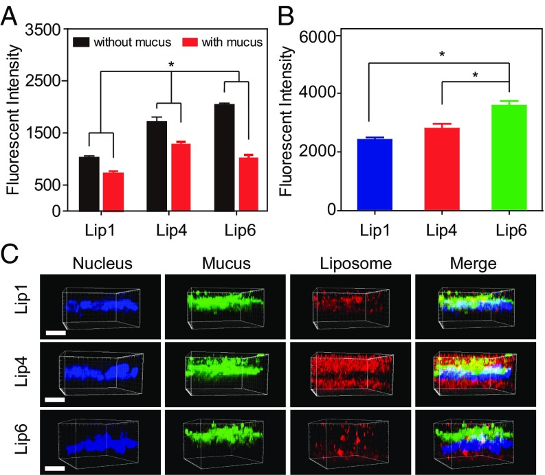
Fig. 8.
Cellular uptake of liposomes in vitro. (A) Internalization of liposomes by E12 cells in the presence or absence of mucus. (B) Internalization of liposomes by non–mucus-producing Caco-2 cells. (C) CLSM 3D images showing liposome localization in E12 cell monolayers. DiI-labeled liposomes are shown in red, mucus stained with Alexa Fluor 488-wheat germ agglutinin (WGA) is shown in green, and cell nuclei stained with Hoechst stain are shown in blue. (Scale bar, 20 μm.) The data are shown as the means ± SD (n = 3). *P < 0.05.

