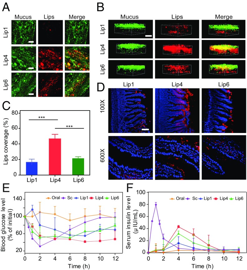
Fig. 9.
Oral delivery of liposomes to rats. The mucus-penetrating ability of liposomes was examined via (A) 2D CSLM scanning and (B) 3D CSLM scanning. To analyze the distribution of liposomes in mucus, Alexa Fluor 488-WGA was used to label the mucin fibers. The DiI-labeled liposomes were then injected into intestinal loops, followed by incubation for 30 min with gentle agitation. (Scale bar, 20 μm.) (C) Quantification of liposome coverage in the mucus shown in A. The data are shown as the means ± SD (n = 3). ***P < 0.001. (D) Distribution of DiI-labeled liposomes in middle intestinal sections at 1 h. Fasted Sprague-Dawley rats were orally administered DiI-labeled liposomes, and the middle intestinal sections were collected for CLSM analysis. Red fluorescence refers to liposomes, and blue fluorescence refers to cell nuclei. (Scale bar, 100 μm.) (E and F) Blood glucose levels (E) and plasma insulin concentrations (F) in diabetic rats following the oral administration of insulin (oral, 30 IU/kg), the s.c. injection of insulin (Sc, 5 IU/kg), and via insulin-loaded liposomes (Lip1, Lip4, and Lip6) (mean ± SD; n = 5).

