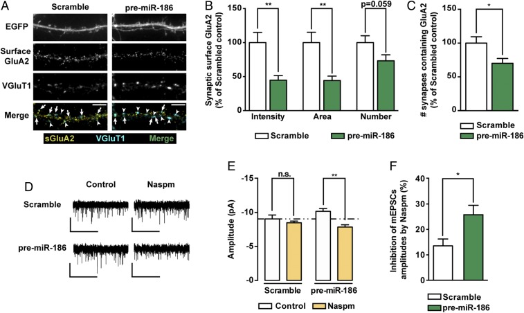
Fig. 5.
Expression of premiR-186 regulates the expression of endogenous GluA2. (A) Representative images of hippocampal neurons expressing premiR-186 or control scramble constructs and stained for surface GluA2, VGluT1, and MAP2. Transfected neurons were identified by expression of EGFP from the premiR-186 expression plasmid. Arrows indicate colocalization of surface GluA2 (yellow) and VGluT1 puncta (cyan); arrowheads pinpoint GluA2-lacking VGluT1 puncta (cyan). (Scale bars, 5 μm.) (B) Intensity, area and number of endogenous synaptic surface GluA2 clusters (colocalizing with VGluT1 clusters) normalized to synapse density (n = 29–32 cells per condition from three independent experiments; Mann–Whitney test: **P ≤ 0.01). (C) Neurons expressing premiR-186 displayed a decreased density of VGluT1+ synapses containing surface GluA2 clusters (n = 29–32 cells per condition, three independent experiments; Mann–Whitney test: *P ≤ 0.05). (D) Representative whole-cell current traces of AMPAR-mediated mEPSCs from hippocampal neurons expressing a scramble control or premiR-186, under control conditions or preincubated with 20 μM Naspm for 30 min. (Scale bars: vertical, 10 pA; horizontal, 5 s.) (E) Naspm decreased the amplitude of AMPAR-mediated mEPSCs of neurons expressing premiR-186 (n = 7–10 cells per condition, five independent experiments; two-way ANOVA with Tukey’s multiple comparison test: **P ≤ 0.01). (F) PremiR-186 expressing neurons displayed a higher percentage of inhibition with Naspm than control neurons (n = 7–9 cells per condition, five independent experiments; Mann–Whitney test: *P ≤ 0.05).

