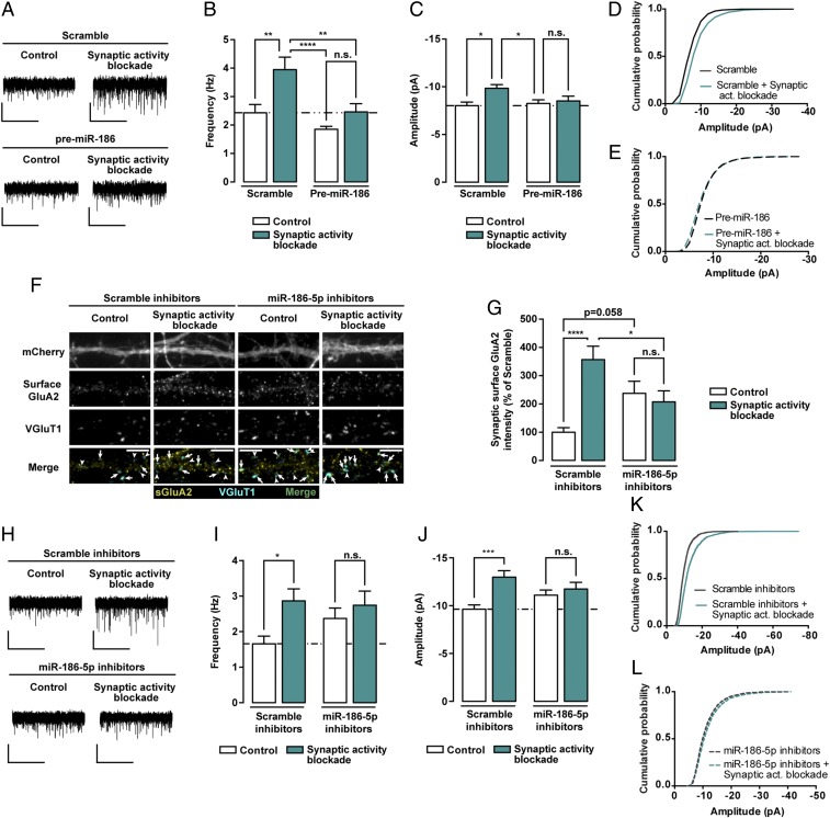
Fig. 7.
Manipulation of basal miR-186-5p levels blocks synaptic scaling-up. (A) Representative whole-cell current traces of AMPAR-mediated mEPSCs from hippocampal neurons expressing a scramble control or premiR-186, and treated with 50 μM GYKI-52466 and MK-801 for 24 h. (Scale bars: vertical, 10 pA; horizontal, 5 s.) (B and C) Expression of premiR-186 hinders the increase of AMPAR-mediated mEPSC frequency (B) and amplitude (C) induced by chronic blockade of synaptic activity (n = 12–14 neurons per condition, six independent preparations). (D and E) Cumulative probability histograms of mEPSC amplitudes display the synaptic scaling associated with chronic blockade of excitatory synaptic activity in neurons expressing scramble control (D), but not in premiR-186–expressing neurons (E) (n = 1,800 events recorded from 16 cells per condition, six independent experiments). (F) Representative images of hippocampal neurons expressing either miR-186-5p inhibitors or control scramble constructs, in control conditions or treated with GYKI-52466 and MK-801 for 24 h, and stained for surface GluA2, VGluT1, and MAP2. Transfected neurons were identified by expression of mCherry from the bicistronic miR-186-5p inhibitors expression plasmid. Arrows indicate colocalization of surface GluA2 (yellow) and VGluT1 puncta (cyan); arrowheads pinpoint GluA2-lacking VGluT1 puncta (cyan). (Scale bars, 5 μm.) (G) Inhibition of basal miR-186-5p expression occluded upscaling of synaptic GluA2 clusters intensity upon blockade of synaptic activity. Synaptic GluA2 clusters (colocalized with VGluT1) were normalized to synapse density (n = 29–30 neurons per condition, three independent experiments). (H) Representative whole-cell current traces of AMPAR-mediated mEPSCs from hippocampal neurons expressing a scrambled sequence or miR-186-5p inhibitors, and exposed to GYKI-52466 and MK-801 for 24 h. (Scale bars: vertical, 10 pA; horizontal, 5 s.) (I and J) Inhibiting the basal levels of miR-186-5p hampered the increase of AMPAR-mediated mEPSC frequency (I) and amplitude (J) induced by blockade of synaptic activity (n = 16–18 neurons per condition, 10 independent preparations). (K and L) Cumulative probability histograms of mEPSC amplitudes display synaptic scaling associated with chronic synaptic activity suppression in neurons expressing scrambled inhibitors (K), but not in miR-186-5p inhibitor-expressing neurons (L) (n = 2,400 events, 16 cells per condition, 10 independent experiments). (B, C, G, I, and J) Statistical analysis was performed using two-way ANOVA with Tukey’s multiple comparison test: *P ≤ 0.05, **P ≤ 0.01, ***P ≤ 0.001, ****P ≤ 0.0001.

