Fig. 2.
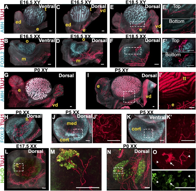
Innervation invades the interior of the ovary but not the testis. (A–D) E16.5 XY (A and C) or XX (B and D) gonads imaged from the ventral (A and B) or dorsal (C and D) side. (E and F) E18.5 XY (E) or XX (F) gonads imaged from the dorsal side. E′ and F′ are magnified views of the areas outlined in E and F that represent the Top and Bottom (28 µm deeper) optical sections from the Z-stacks used to generate maximum-intensity projections shown in E and F. (G and H) P0 XY (G) or XX (H) gonads imaged from the dorsal side. (I–K) P5 XY (I) or XX (J and K) gonads imaged from the dorsal (I and J) or the ventral side (K). I′, J′, and K′ are magnified views of TUJ1 staining from the areas outlined in I, J, and K. Note that TUJ1 also stains Sertoli cells at some stages (I and I′). (L–O) E17.5 (L and M) and P0 (N and O) XX gonads stained for the pan-neuronal marker TUJ1 (red, A–O) and neural body marker HuC/D (green, L–O). M and O are higher-magnification images of the areas outlined in L and N. Note that HuC/D also stains oocytes (L–O and SI Appendix, Fig. S3). HuC/D staining in the oocyte is distinct from the stain observed in neural bodies: white arrowheads in O point to TUJ1-/HuC/D+ oocytes, and white arrows in O point to TUJ1+/HuC/D+ neural cell bodies. XY samples were stained for the Sertoli cell marker AMH (cyan, A, C, E, G, and I), and XX samples were stained for the granulosa cell marker FOXL2 (cyan, B, D, F, H, J, and K). All samples were counterstained with Hoechst nuclear dye (grayscale). cort, cortex; e, epididymis; ed, efferent ductules; h, hilus; m, mesonephros; med, medulla; o, ovary; t, testis; tt, testis tubules; vd, vas deferens. (Scale bars, 100 µm.)

