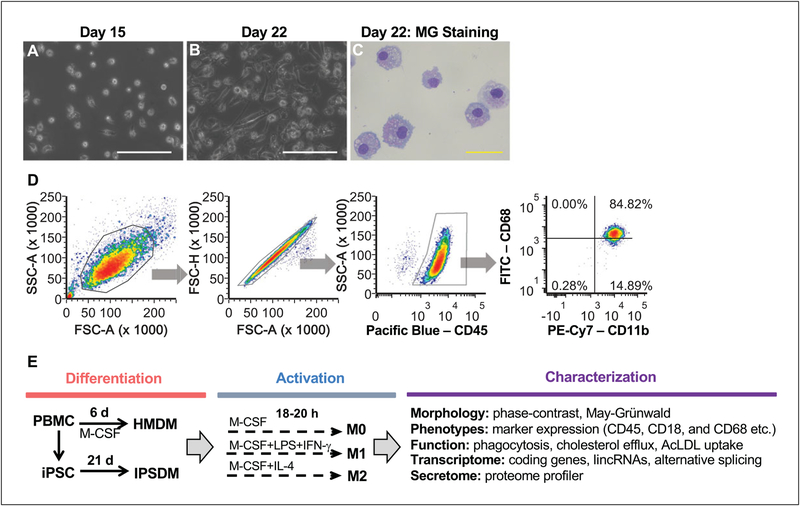
Figure 3.
The morphology and marker expression of IPSDM. (A) The seeding density of day 15 myeloid progenitors harvested from the suspension culture of EBs. (B) The phase-contrast image of day 22 IPSDM. The white bar = 100 μm. (C) May-Grünwald Giemsa (MG) staining of day 22 IPSDM showing macrophage-like morphology. The yellow bar = 50 μm. (D) Macrophage marker expression of day 22 IPSDM characterized by FACS. The day 22 IPSDM are CD45+/CD18+/CD11b+ with high expression of CD68. Additional morphological, phenotypical, and functional characterizations as outlined in (E) were performed and the results can be found in our previous publication (Zhang et al., 2015; Zhang, Shi et al., 2017; Zhang, Xue et al., 2017).

