
Figure 2. ALM and thalamus show similar neural dynamics.
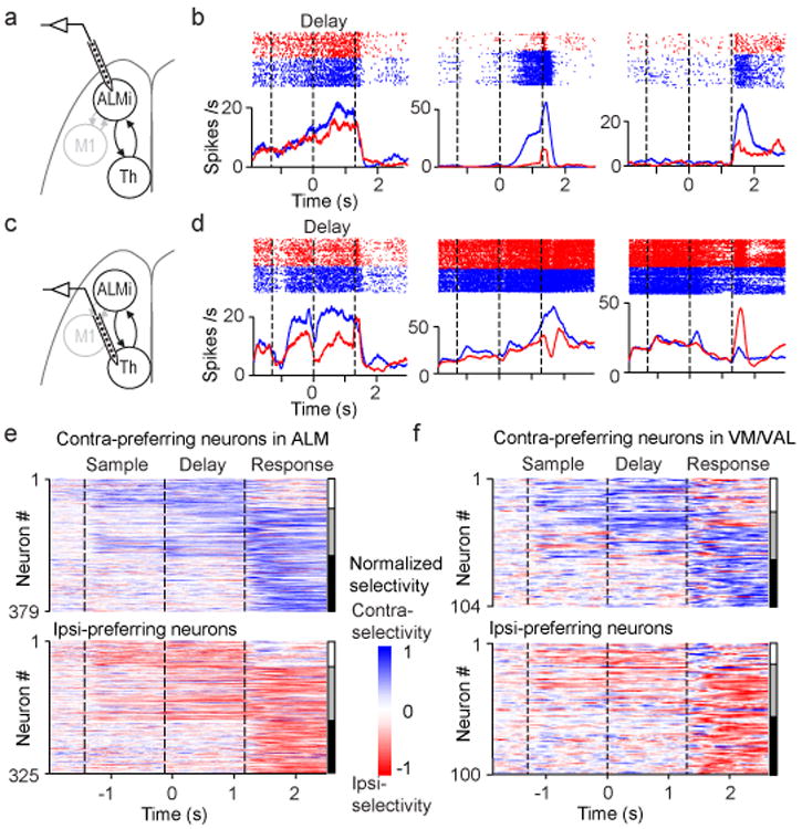
a. Silicon probe recordings in ALM (relevant to b, e).
b. Three example ALM neurons. Top, spike raster. Bottom, peri-stimulus time histogram. Blue, correct contra trials; red, correct ipsi trials. Dashed lines separate behavioral epochs.
c. Silicon probe recordings in VM/VAL (relevant to d, f).
d. Three example VM/VAL neurons. Same format as b.
e. ALM population selectivity (n = 704). Vertical bars on the right; white, neurons with preparatory activity only; grey, both preparatory activity and peri-movement activity; black, peri-movement activity.
f. VM/VALpopulation selectivity (n = 204). Same format as e.