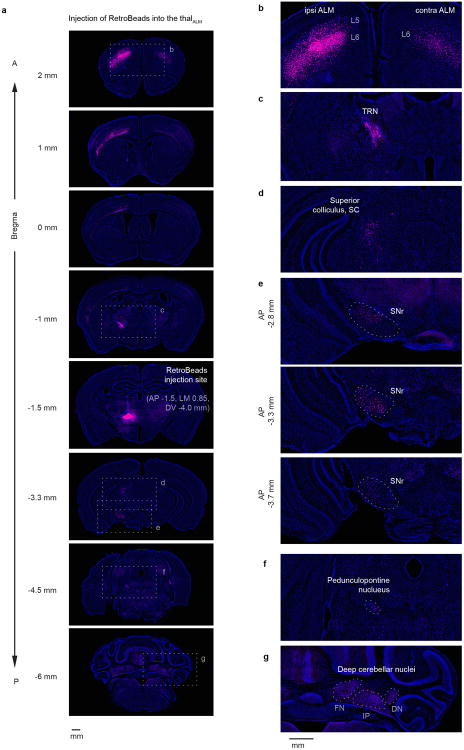
Extended Data Figure 9. Thalamic regions that are connected reciprocally with ALM (thalALM) receive input from multiple brain areas.
RetroBeads were injected into thalALM (AP -1.5, ML 0.85, DV -4.0 mm from Bregma, mainly in VM). Magenta, retrograde labeling; blue, Nissl staining.
a. Coronal sections. Dashed boxes indicate location of magnified images on right panels (b-g).
b. Labeling in ALM. Overall labeling was much stronger in ipsilateral ALM. Labeling in ALM was observed on both sides in L6, whereas labeling in L5 was seen only in ipsilateral ALM. L6 neurons are corticothalamic neurons, whereas the L5 neurons correspond to pyramidal tract neurons that send a branch to thalamus60. In addition to ALM, labeling was observed in M1, S2 and weakly in other cortical areas (see a).
c. Labeling in the ipsilateral thalamic reticular nucleus, TRN.
d. Labeling in the ipsilateral superior colliculus, SC.
e. Labeling in the ipsilateral substantia nigra pars reticulate (SNr), in three coronal sections. Labeling was observed throughout SNr from the caudal to the rostral end, consistent with a previous report54.
f. Labeling in the ipsilateral pedunculopontine nucleus (PPN).
g. Labeling in the contralateral deep cerebellar nuclei. FN, fastigial nucleus; IP, interposed nucleus; DN, dentate nucleus.

