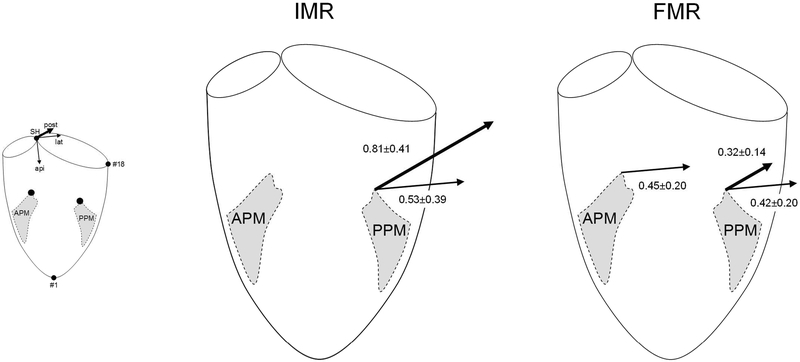
Figure 1:
Schematic illustrations depicting three-dimensional anterior and posterior papillary muscle (APM and PPM, respectively) displacement vectors in experimental ovine models of ischemic and functional mitral regurgitation (IMR and FMR, respectively). Arrows indicate vectors that reached statistical significance according to Table 2. Arrow lengths are proportionate to the average of the differences between Control and the respective IMR/FMR values. The small schematic illustrates the coordinate system used (see Methods). SH=saddle horn, api=apical, lat=lateral, post=posterior, #18=mid-lateral mitral annular marker.

