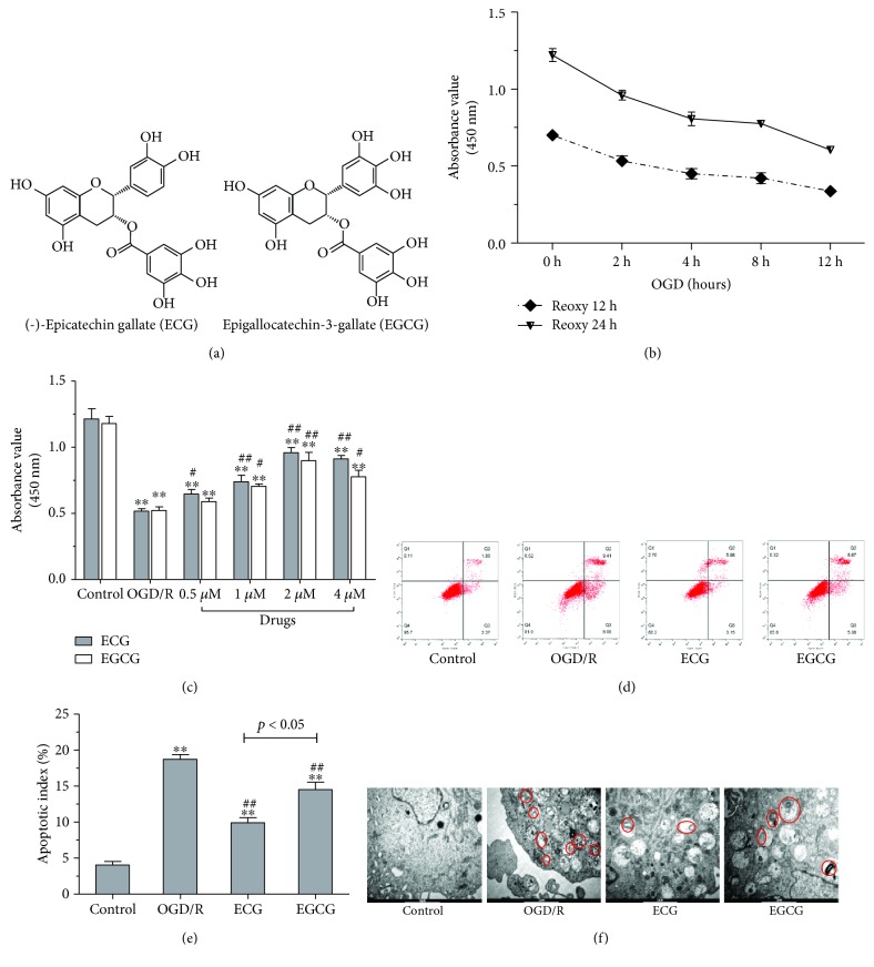
Figure 1.
Effect of ECG/EGCG on cell viability, apoptosis, and autophagy in HBMVECs of OGD/R. Chemical structures of ECG and EGCG are shown in (a). Cell viability was assayed by CCK-8 when HBMVECs were cultured in glucose-free culture and oxygen deprivation of 0.5% O2 + 5% CO2 + 94.5% N2 for 2 h, 4 h, 8 h, or 12 h and then reoxygenated for 12 or 24 h in normal medium (b). Cell viability of HBMVECs treated with 0.5, 1, 2, and 4 μM of ECG/EGCG in OGD/R measured by CCK-8 (c). Effect of ECG/EGCG on apoptosis induced by OGD/R, measured by flow cytometry, was explored (d and e). Autophagy was also observed using electron microscopy (f). Data was given as mean ± SD, n = 6. ∗ P < 0.05 and ∗∗ P < 0.01 compared with control, # P < 0.05 and ## P < 0.01 versus cells after OGD/R treatment with solvent (OGD/R or solvent control group).

