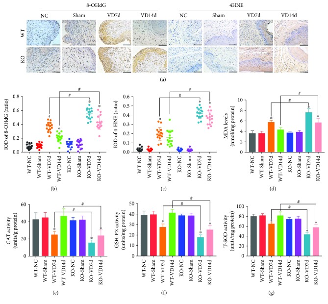
Figure 4.
Oxidative damage and antioxidative capacity in the anterior wall of mice. (a–c) Immunohistochemistry was used to detect the levels of 8-OHdG and 4-HNE in the anterior wall of mice in eight groups. Brown represents the positive signal. Scale bars: 100 μm. (d) The MDA measurement kit was used to detect the levels of MDA in the anterior wall of mice in eight groups. (e–g) CAT, GSH-PX, and T-SOD measurement kits were used to determine the activities of CAT, GSH-PX, and T-SOD, respectively, in the anterior wall of mice in eight groups. ∗ P < 0.05 compared with the WT-VD group, # P < 0.05 between two groups. Every experiment was repeated 3 times and representative figures are presented here. 8-OHdG: 8-oxo-2′-deoxyguanosine; 4-HNE: 4-hydroxynonenal; MDA: malondialdehyde; CAT: catalase; GSH-PX: glutathione peroxidase; T-SOD: total superoxide dismutase; VD: vaginal distension; WT-NC: nonoperated control group of wild-type mice; WT-Sham: sham-operated group of wild-type mice; WT-VD7d: VD group of wild-type mice and evaluated on day 7 after VD; WT-VD14d: VD group of wild-type mice and evaluated on day 14 after VD; KO-NC: nonoperated control group of Nrf2-knockout mice; KO-Sham: sham-operated group of Nrf2-knockout mice; KO-VD7d: VD group of Nrf2-knockout mice and evaluated on day 7 after VD; KO-VD14d: VD group of Nrf2-knockout mice and evaluated on day 14 after VD.

