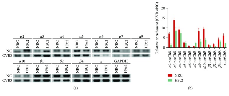
Figure 1.
The mRNA expression of nAChRs in CVB3-infected NRC and H9c2 cells. NRC and H9c2 cells were cultured for 48 h in 10% FBS medium and then stimulated by CVB3 for 2 h. A continuous culture for 24 h was accepted after CVB3 was removed and total mRNA was then collected. PCR for GAPDH was regarded as the loading control. (a) RT-PCR showing the mRNA expression of nAChR subunits in NRC and H9c2 cells with or without CVB3 infection. (b) RT-qPCR showing the fold change of the mRNA expression of nAChR subunits in CVB3-infected NRC and H9c2 cells compared to normal controls.

