
Figure 3.
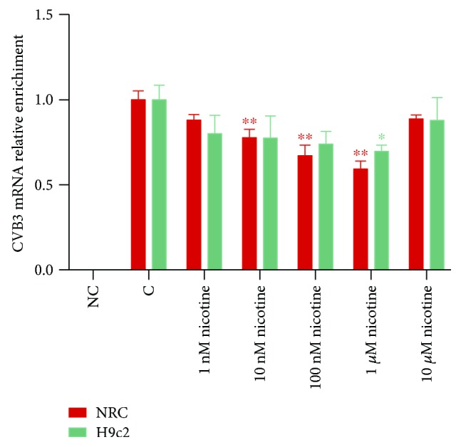
The mRNA expression of CVB3 in NRC and H9c2 cells after treatment with different concentrations of nicotine. After being infected by CVB3 for 2 h, the culture medium was removed and different concentrations of freshly dissolved nicotine were added to stimulate nAChRs. Total mRNA was collected 36 h later for the evaluation of CVB3 mRNA by RT-qPCR. ∗P < 0.05 and ∗∗P < 0.01 vs. the control group.