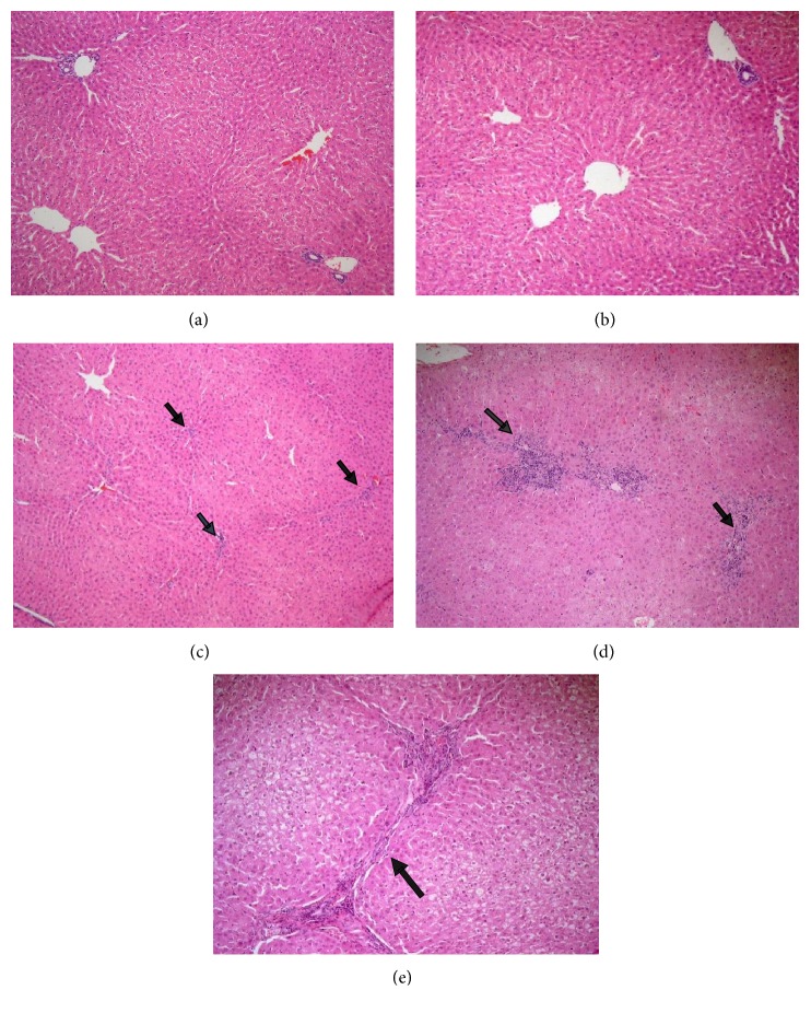
Figure 6.
Effect of LSS on histopathological changes of liver tissues of normal groups in rabbits. (a) Showing normal histological picture of hepatic lobule that consists of central vein surrounded by normal hepatocytes (HEx100). (b) Showing liver sections of the rabbits treated with LSS (200 mg/kg bw.) which revealed normal histological picture of hepatic lobule that consists of central vein surrounded by normal hepatocytes (HEx100). (c) Showing liver sections of the rabbits treated with LSS (400 mg/kg bw.) which revealed normal hepatic architecture with only mild inflammation [arrows] (HEx100). (d) Showing liver sections of the rabbits treated with LSS (400 mg/kg bw.) which revealed evident chronic venous congestion and moderate inflammation [arrows] (HEx100). (e) Showing liver sections of the rabbits treated with LSS (400 mg/kg bw.) which revealed portal to portal bridging fibrosis [arrow] (HEx200).

