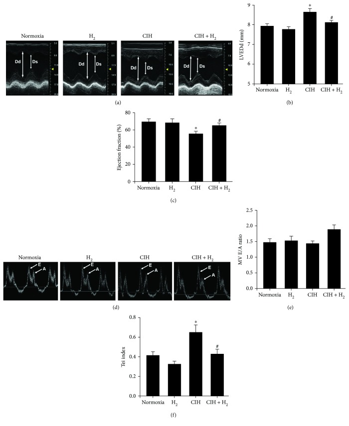
Figure 1.
The effect of H2-O2 mixture on improving cardiac dysfunction in a rat model when exposed to CIH for 35 d. (a) M-model echocardiography of the short axial section of the thoracic bones in rats. Dd: end-diastolic diameter of the left ventricle; Ds: end-systolic diameter of the left ventricle. (b) The mean value of the left ventricular end-diastolic internal diameter (LVEDd). (c) The ejection fraction (EF) of the left ventricle. (d) Rat four-chamber echocardiograph with atrial contraction waves. (e) The velocity ratio of the E peak to the A peak in the cardiac mitral valve (MV E/A); (f) Tei index = (IVCT + IVRT)/ET. ∗p < 0.05 vs. normoxia group; #p < 0.05 vs. CIH group; n = 5.

