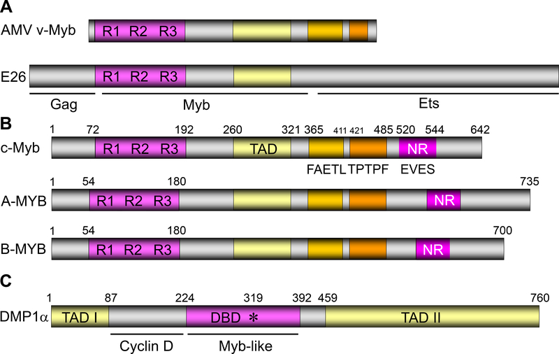
Figure 1. The domain structures of Myb-like proteins.
A. The structure of AMV and E26 proteins that encode v-Myb. R1–3 represents the three repeats within the Myb protein responsible for DNA binding. See refs. 1–3, 105–111.
B. The domain structures of A-Myb, B-Myb, and c-Myb (53, 170). TAD: transactivation domain; NR: negative regulatory domain. The “FAETL” domain (171) is required for oncogenic activity, the “TPTPF” domain conserved in the other Myb proteins, and the “EVES” domain (172) that is involved in intra‑molecular interactions and negative regulation.
C. The domain structure of the human DMTF1α protein (58). DBD: DNA-binding domain. This protein has three tandem MYB-like repeats and two transactivation domains.

