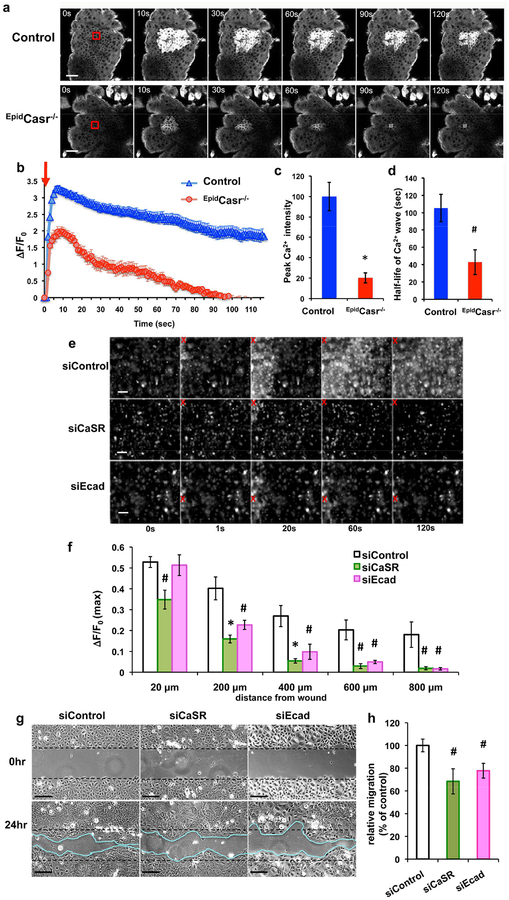
Figure 3. Blocking CaSR or E-cadherin expression diminished wound-induced Ca2+i propagation and collective keratinocyte migration.
(a-d) Dorsal skins excised from EpidCasr−/−//GCaMP+/+ (EpidCasr−/−) and EpidCasr+/+//GCaMP+/+ (control) mice were subjected to laser irradiation in the stratum basale (SB). (a) Time-lapse GCaMP fluorescent images of SB and the quantitative measurements of (b) magnitude, (c) peak intensity, and (d) duration of the Ca2+i propagation following wounding. Mean+/−SE (n=35–60 cells), * P<0.01, # P<0.05. (e-h) Human keratinocytes were transfected with scrambled siRNA (siControl) or specific siRNA targeting CaSR (siCaSR) or E-cadherin (siEcad) prior to scratch wounding. (e, f) Confluent cultures were loaded with calcium green before imaging. (e) Time-lapse fluorescent images and (f) quantitation of temporal changes in the intensity (peak ΔF/F0) of Ca2+i propagations at various distances from wound site. Red X indicates wounded sites. Data were presented as mean+/−SE of 6 separate experiments. *P<0.01, #P<0.05. (g) Representative micrographs of keratinocyte sheets at 0 and 24hr after scratch wounding. Bar = 50 (a, g) or 100 μm (e). (h) Scratched areas re-occupied by migrating keratinocytes were quantified 24hr after wounding and normalized to siControl, (n=14), # P<0.05.

