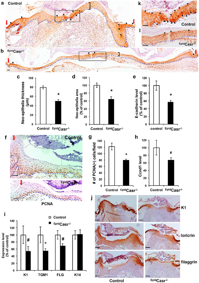
Figure 4. CaSR ablation reduced E-cadherin expression, keratinocyte proliferation, and differentiation in the neo-epithelia.
Skin wounds were excised from EpidCasr−/− mice and control littermates (a-h, k, l) four or (i-j) five days after injury, sectioned, and stained with antibodies against (a, b) E-cadherin, (f) PCNA, or (j) differentiation markers. Basement membranes are outlined with dotted blue lines. Red arrows denote the wound margins and black brackets mark the spans of neo-epithelia. Arrowheads indicate areas with the strongest staining of E-cadherin in the cell membrane. Boxed areas in a and b are enlarged and shown in panel k and l, respectively. Bar = 50 (a, b, f, k, l) or 100 m (j). The (c) thickness and (d) tissue size of neo-epithelia and (g) the number of PCNA(+) cells in representative fields were quantified and presented as mean+/−SE (n=16). Expression of E-cadherin (e), cyclin D1 (h) and differentiation markers (i) in wounds were determined by qPCR and normalized to the levels in control mice (n=6–8); * P<0.01, # P<0.05.

