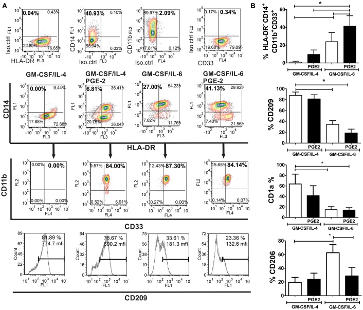
Figure 1.
Phenotypic analysis of M-MDSC and DC generated from human monocytes in vitro. (A,B) The monocytes were cultivated in the presence of GM-CSF/IL-4, GM-CSF/IL-4/PGE2, GM-CSF/IL-6, or GM-CSF/IL-6/PGE2 for 5 days, followed by their phenotypic analysis. (A) A representative analysis of the co-expression of HLA-DR, CD14, CD11b, and CD33 is shown. The doublets and the dead (FSClow) cells were gated-out (not shown), and the quadrants were set according to the single-labeled samples (first row). CD11b/CD33 plots were gated from the HLA-DR−CD14+ region, and the percentage of HLA-DR−CD14+CD33+CD11b+ was calculated based on these plots. The expression of CD209 was analyzed within the total gated cell population. (B) The summarized results on % of HLA-DR−CD14+CD33+CD11b+ cells, CD209+, CD1a+, and CD206+ are shown as mean ± SD from 4 independent experiments carried out with different donors. *p < 0.05 between the indicated samples (RM ANOVA, Tukey post-test).

