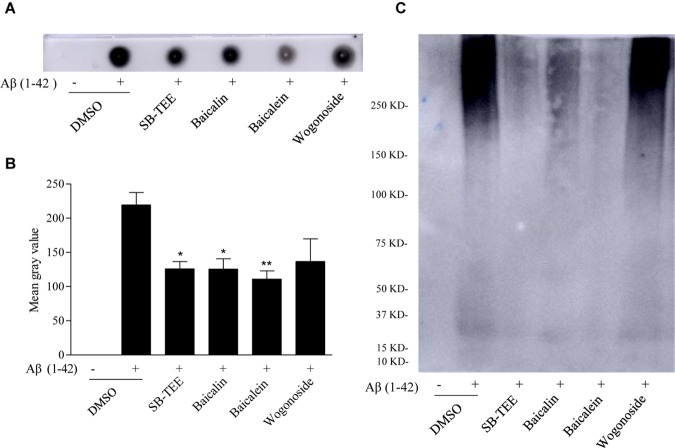
FIGURE 5.
Inhibitory effect of SB-TEE and selected compounds on Aβ (1–42) fibrillation by dot blot assay and native gel electrophoresis analysis. (A) The dot blot image of Aβ (1–42) with or without the incubation with SB-TEE (100 μg/mL), baicalin (50 μM), baicalein (50 μM), or wogonoside (50 μM). (B) The dot blot images were analyzed and quantitated with the software Image J. Columns, means of 3 independent experiments; bars, SEM. ∗p < 0.05, ∗∗p < 0.01 vs. 20 μM Aβ (1–42) alone group. (C) The native gel electrophoresis analysis was performed by analyzing Aβ (1–42) solution (50 μg) with or without incubation of SB-TEE, baicalin, baicalein or wogonoside at 37°C for 5 days using the 4–16% gradient native gel under non-denatured condition. The full-length images of dot blot and native gel electrophoresis are displayed in Supplementary Figure S1.

