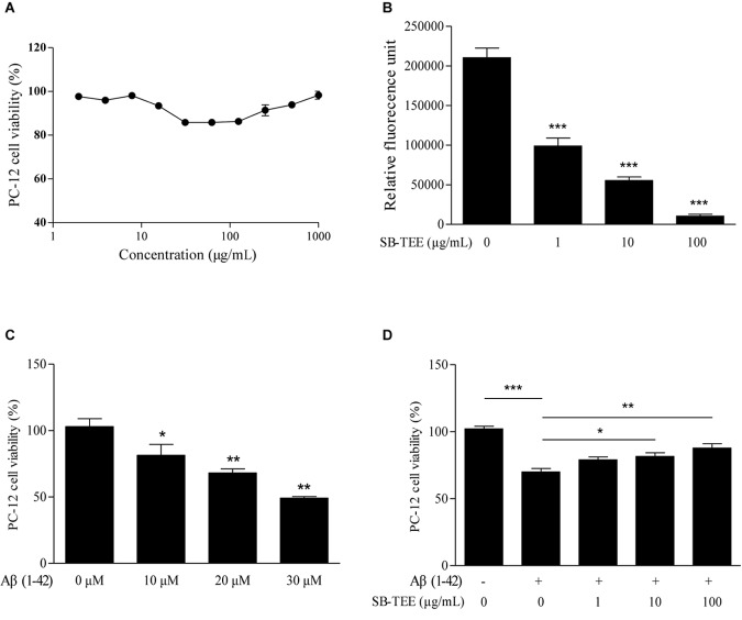
FIGURE 6.
Cytotoxicity of SB-TEE and Aβ (1–42) fibril on PC-12 cells. (A) Cytotoxicity of SB-TEE on PC-12 was examined by MTT assay at 48 h after treatment. (B) The anti- Aβ fibrillation effect of SB-TEE (1–100 μg/mL) was measured by ThT fluorescence assay. ∗∗∗p < 0.001 vs. 20 μM Aβ (1–42) group. (C) Cellular toxicity induced by 10–30 μM of Aβ (1–42) on PC-12 cells was evaluated by MTT assay at 48 h after treatment. ∗p < 0.05 and ∗∗p < 0.01 vs. 0 μM Aβ (1–42) group. (D) Cellular toxicity induced by 20 μM of Aβ (1-42) on PC-12 cells was evaluated by MTT assay at 48 h after SB-TEE (1, 10, and 100 μg/mL) treatments. Column, means of 3 independent experiments; bars, SEM. ∗p < 0.05, ∗∗p < 0.01 and ∗∗∗p < 0.001 vs. 20 μM Aβ (1–42) alone group.

