
Figure 5.
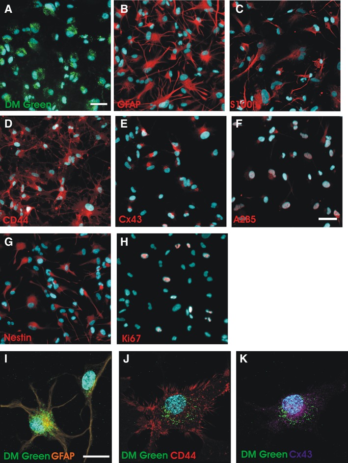
Immunohistochemical analysis of Q‐Cells after labeling with CS‐1000 DM green (A) Q‐Cells were incubated with 1 mg/ml CS‐1000 DM green for 1 week followed by incubation with 10% fetal bovine serum (FBS) for 96 hours (scale bar = 50 μm). (B–E): Q‐Cells differentiate into astrocytes and express appropriate astrocytic markers. (F, G): A2B5 and nestin immunostaining is still present after 4 days of FBS exposure suggesting a transition from progenitor to more mature astrocyte identity. Scale bar = 50 μm. (H): Few Ki67+ phenotypes are observed in differentiated Q‐Cells. (I–K): Confocal microscopy shows perinuclear DM green localization in differentiating astrocytes. Scale bar = 25 μm.