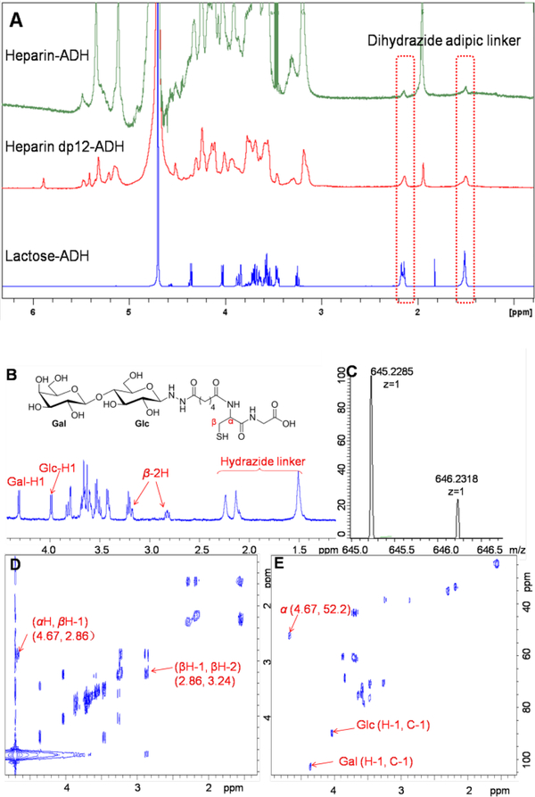
Fig. 1.
NMR and HRMS analysis. Panel (A) shows 1D 1H NMR spectra of heparin-, heparin dodecasaccharide- and lactose-ADH. Two sets of peaks at 1.51 and 2.16 ppm in the 1H NMR correspond to dihydrazide adipic linker. Panel (B), (D), (E) and (C) show the 1D 1H, 2D 1H-1H COSY, 1H-13C HSQC NMR and HRMS (positive-mode) spectra of lactose-dipeptide (Cys-Gly) conjugate.

