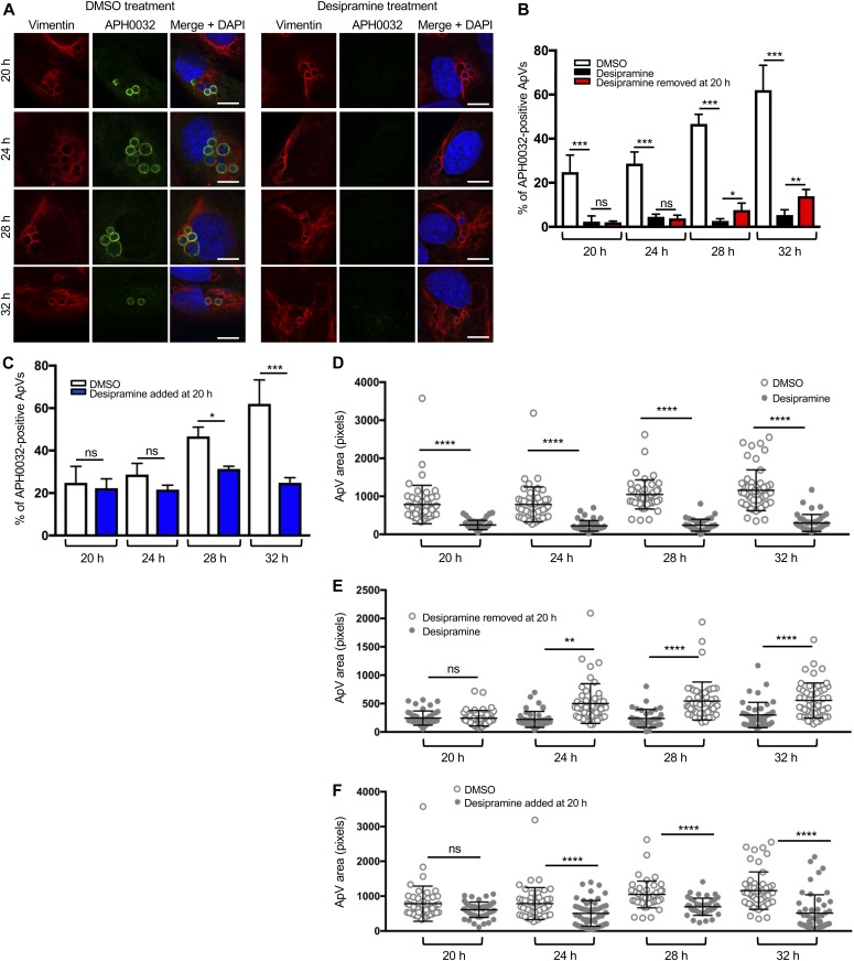
Figure 2. Functional inhibition of ASM halts ApV maturation and expansion.
(A, B, D, E) Desipramine was added to RF/6A cells before infection with A. phagocytophilum and treatment was either maintained throughout the time course (A, B, D) or removed at 20 h (B, E). (C, F) Desipramine was added to A. phagocytophilum–infected RF/6A cells beginning at 20 h (C, F). DMSO served as vehicle control. At 20, 24, 28, and 32 h, the cells were fixed and examined by confocal microscopy for ApV maturation (A–C) and expansion (D–F). (A–C) Desipramine reversibly inhibits APH0032 expression and localization to the ApV. A. phagocytophilum–infected RF/6A cells were screened with antibodies targeting vimentin and APH0032 to demarcate and assess maturation of the ApV, respectively. DAPI was used to stain host cell nuclei and bacterial DNA. (A) Representative confocal micrographs of desipramine or DMSO-treated cells at the indicated postinfection time points. (B, C) Percentages of APH0032-positive ApVs determined for 100 cells for each of three biological replicates per condition. (D–F) Desipramine reversibly inhibits ApV expansion. The mean ApV pixel area was determined for 50 ApVs per time point per condition. Error bars indicate SD. t test was used to test for a significant difference among pairs. Statistically significant (*P < 0.05; **P < 0.01; ***P < 0.001; ****P < 0.0001) values are indicated. ns, not significant. Data shown are representative of three experiments conducted in triplicate with similar results. Scale bar = 10 μm.

