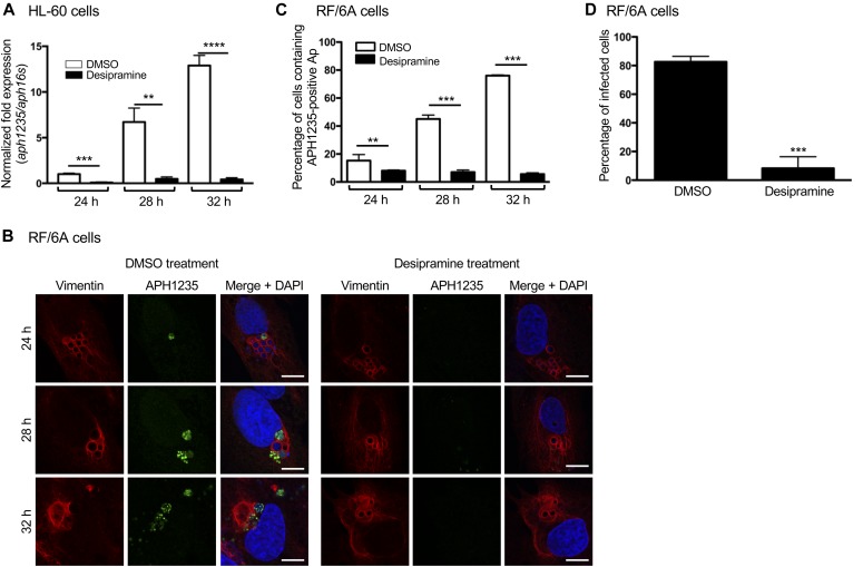
Figure 3. Desipramine inhibits A. phagocytophilum conversion to the infectious form.
(A) Desipramine inhibits aph1235 transcription. Desipramine-treated HL-60 cells were infected with A. phagocytophilum. Total RNA isolated at 24, 28, and 32 h was subjected to qRT-PCR. The 2−ΔΔCT method was used to determine the relative aph1235 expression level normalized to that of A. phagocytophilum 16S rRNA. (B, C) Desipramine inhibits APH1235 protein expression. Desipramine-treated RF/6A cells were infected with A. phagocytophilum. At 24, 28, and 32 h, the cells were fixed, immunolabeled with APH1235 and vimentin antibodies, stained with DAPI, and visualized using confocal microscopy. (B) Representative confocal micrographs. Scale bar = 10 μM. (C) Percentage of APH1235-positive ApVs determined by counting 100 cells for each of triplicate samples per time point. (D) Desipramine inhibits A. phagocytophilum–infectious progeny production. RF/6A cells were treated with desipramine or DMSO followed by infection with A. phagocytophilum. At 48 h, the cells were mechanically disrupted followed by isolation and subsequent incubation of host cell–free bacteria with naïve untreated cells. At 24 h, the recipient cells were fixed and examined by immunofluorescence microscopy to determine the percentage that had become infected. Error bars indicate SD. t test was used to test for a significant difference among pairs. Statistically significant (**P < 0.01; ***P < 0.001; ****P < 0.0001) values are indicated. Data shown in (A–C) are representative of three experiments conducted in triplicate that yielded similar results. Data shown in (D) are representative of two experiments conducted in triplicate with similar results.

