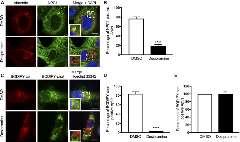
Figure 4. Desipramine alters NPC1-mediated cholesterol trafficking to the ApV.
Desipramine treatment inhibits NPC1 localization to the ApV. Desipramine- or DMSO-treated RF/6A cells were infected A. phagocytophilum. (A–E) At 24 h, the cells were either fixed, immunolabeled with vimentin and NPC1 antibodies, stained with DAPI, and examined by confocal microscopy (A, B); or incubated with BODIPY ceramide (cer) or BODIPY cholesterol (chol), stained with Hoechst 33342, and visualized by live cell imaging (C–E). (A) Representative confocal micrographs of infected cells immunolabeled for vimentin and NPC1. Regions that are demarcated by hatched-line boxes are magnified in the inset panels. (B) Percentage of vimentin-positive ApVs to which NPC1 immunosignal localizes in DMSO- and desipramine-treated cells determined by counting 100 cells for each of triplicate samples per condition. (C) Representative live cell images of infected cells incubated with BODIPY-cer and BODIPY-chol. (D, E) Percentages of ApVs to which BODIPY-cer–positive (D) or BODIPY-chol–positive (E) vesicles localize. Error bars indicate SD. t test was used to test for a significant difference among pairs. Statistically significant (****P < 0.001) values are indicated. ns, not significant. Data shown are representative of three experiments conducted in triplicate that yielded similar results. Scale bar = 10 μm.

