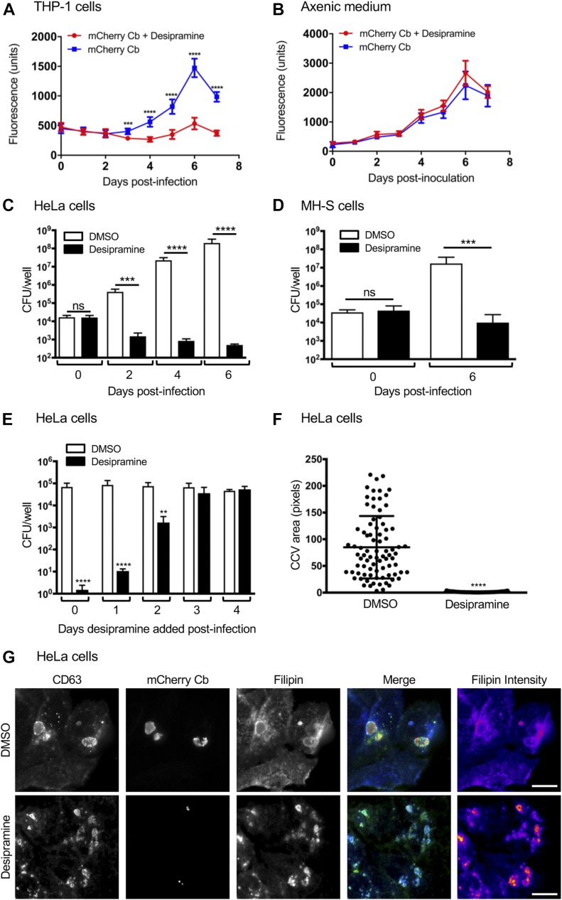
Figure 6. Desipramine-induced cholesterol accumulation in the C. burnetii vacuole is bactericidal.
(A, B) mCherry-C. burnetii (Cb)–infected THP-1 macrophage-like cells (A) or mCherry-C. burnetii grown in axenic medium (B) were treated with desipramine or not treated with. The bacterial load was measured as relative fluorescent units. (C–E) C. burnetii was added to HeLa cells (C, E) or MH-S cells (D) that had been pretreated with desipramine or DMSO, or C. burnetii–infected cells were treated at the indicated days postinfection (E). Bacterial load was measured using a CFU assay. (F) CCV area was determined for desipramine and DMSO-treated C. burnetii–infected HeLa cells. (G) HeLa cells that had been treated with desipramine and infected with mCherry-C. burnetii were labeled with filipin and CD63 antibody. Error bars indicate SD. t test was used to test for a significant difference among pairs. Statistically significant (**P < 0.01; ***P < 0.001; ****P < 0.0001) values are indicated. ns, not significant. Data in panels A and B are representative of three experiments conducted in triplicate with similar results. Data in panels C through F are the means ± SD of three independent experiments. Scale bar = 50 μm.

