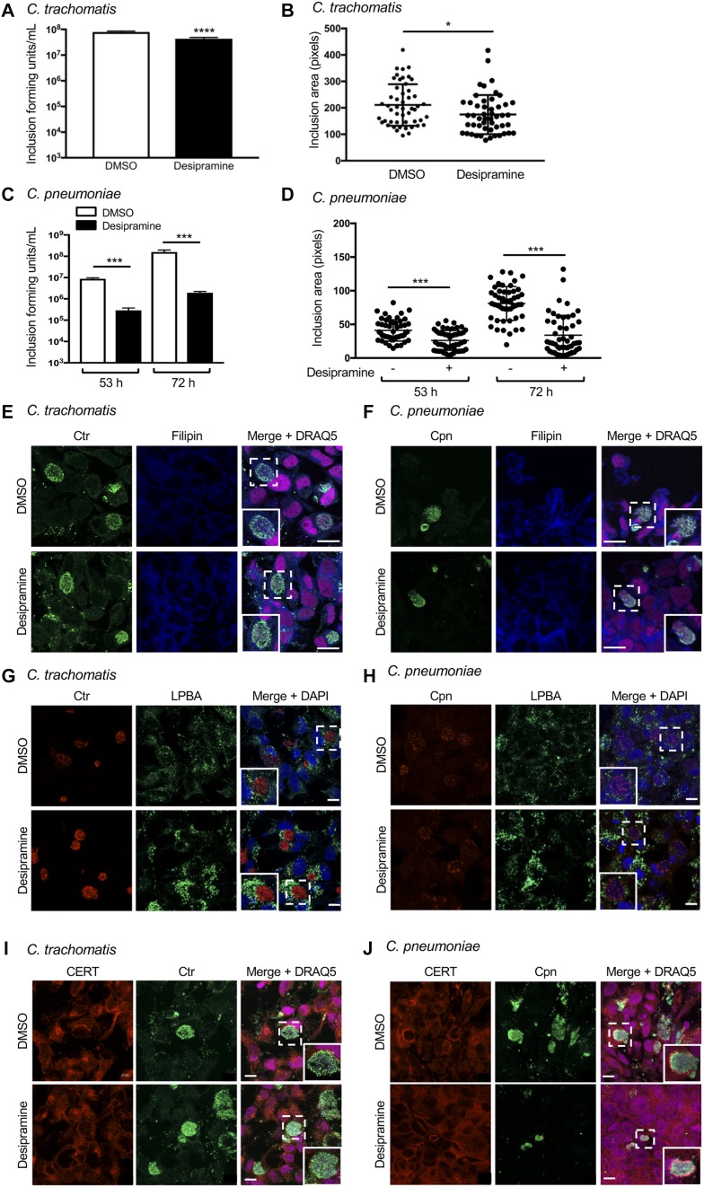
Figure 7. C. trachomatis and C. pneumoniae exhibit reduced FIASMA sensitivity.
(A–D) Desipramine or DMSO-treated HeLa cells infected with C. trachomatis (Ctr; A, B) or C. pneumoniae (Cpn; C, D) were either lysed to recover infectious progeny that were incubated with naïve HeLa cells to determine inclusion-forming units (A, C) or were fixed and assessed by immunofluorescence microscopy to determine inclusion area (B, D). (E–J) Desipramine-treated or control cells were infected with Ctr (E, G, I) or Cpn (F, H, J). The cells were either incubated with filipin (E, F) or screened with antibodies specific for LBPA (G, H) or CERT (I, J) together with antisera against C. trachomatis (E, G, I) or C. pneumoniae (F, H, J). DAPI or DRAQ5 was used to stain DNA. Regions that are demarcated by hatched-line boxes are magnified in the inset panels. Error bars indicate SD. t test was used to test for a significant difference among pairs. Statistically significant (*P < 0.05; ***P < 0.001) values are indicated. Data shown are representative of three experiments conducted in triplicate with similar results. Scale bar = 10 μm.

