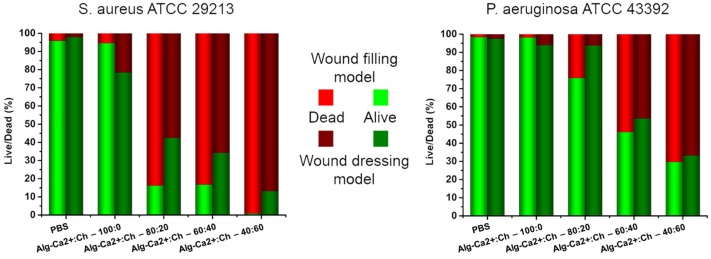
Figure 3.
Bactericidal experiments of chitosan containing hydrogels. Gels with a different positive charge ratio of chitosan and Ca2+ were tested using two different methods. Representative pictures of S. aureus and P. aeruginosa adhered on glass and treated with chitosan containing hydrogels are shown together with a corresponding ratio of dead and alive bacterial cells. Red indicates the amount of dead bacteria while green represents alive bacteria. Light colors show results for direct mixing (wound filling model) and dark colors for the pre-formed gels (wound dressing model). Representative pictures of bacteria can be found in the Supplementary Materials (Figures S3 and S4).

