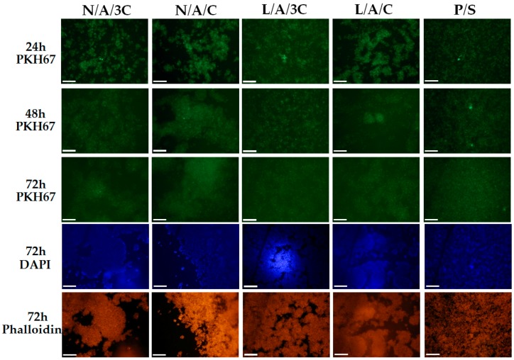
Figure 7.
The morphological changes of RAWs264.7 after 24, 48 and 72 h in the cultures. Cellular membranes were stained with PKH67 (light green), nuclei were visualized with DAPI stating (blue dots), while actin cytoskeleton was visualized using atto-565 phalloidin (red stained cell bodies). Magnification used is 100×, and scale bar is 200 μm.

