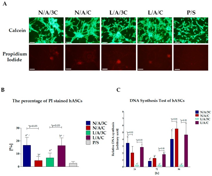
Figure 9.
Cytotoxicity of biomaterials and influence of DNA synthesis in hASC cultures. Images of cells stained with calcein and propidium iodide (A), scale bar =200 μm; Comparative analysis of percentage of propidium iodide positive cells (B); Results of BrdU incorporation assay (C). Fold change in DNA synthesis was calculated by comparing BrdU signals of hASCs maintained with biomaterials to that of the control culture, which was assigned a value of 1. An asterisk (*) indicates a statistically significant difference (p < 0.05) for comparative analysis of changes related to the biomaterial composition, while hashtag (#) indicates differences between cultures on biomaterials and polystyrene.

