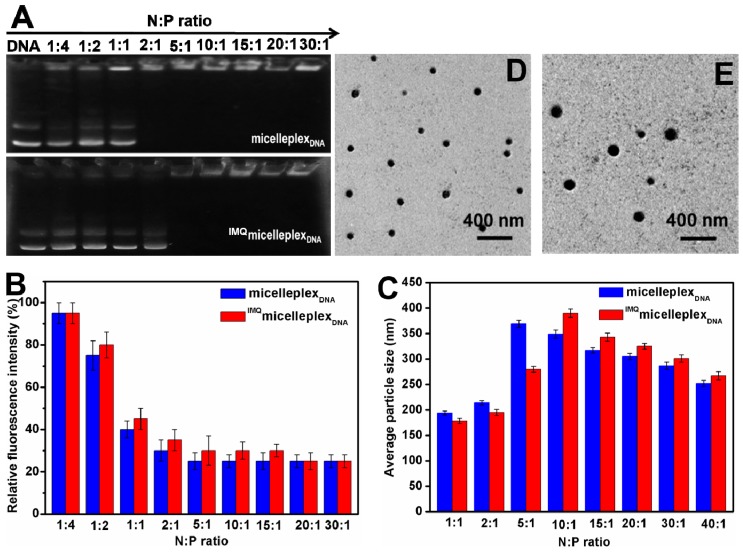
Figure 7.
DNA binding, condensation by micelles, and size and morphology of micelleplexes with or without loading of IMQ. (A) Gel retardation assay; (B) ethidium bromide (EB) exclusion; (C) average particle size by DLS; TEM images of blank micelleplexes (D); and IMQ-loaded micelleplexes (E) at N:P = 20 (The discrete and spherical blackspots represented corresponding micelleplexes).

