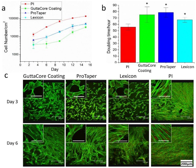
Figure 4.
(a) Proliferation curves of DPSC plated, at an initial cell density of 10,000/cm2 on the Gutta-percha scaffolds (shown in Figure 1) and 25 nm spun cast PI thin films; (b) The doubling time of the DPSC calculated from the data plotted in (a). The Gutta-percha samples differ from control sample at a statistical level of p > 0.05 (*); (c) Confocal microscopy images of DPSC plated on PI and on the three Gutta-percha scaffolds after 3 and 6 days in culture without dexamethasone. Actin filaments were stained with Alex Flour 488 (green) and nucleus was stained with propidium iodide (red). Inset: High magnification images of individual cells in the cultures without dexamethasone (scale bar is 50 μm).

