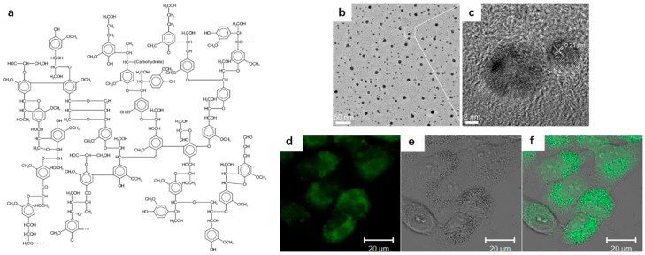
Figure 1.
(a) The molecular structure of lignin; (b) TEM and (c) HRTEM images of CDs; (d) a confocal fluorescence microphotograph of Hela cells labeled with the CDs (λ ex: 405 nm); (e) a bright field microphotograph of the cells; and (f) an overlay image of (d,e). Figure adapted from Ref. [42] with permissions from the publishers.

