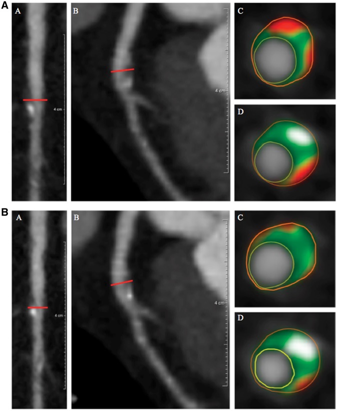
Figure 2.
Left anterior descending artery plaque identified before (2A) and after (2B) biologic therapy. (A) (a) Longitudinal planar and (b) curved planar reformat. (c and d) Representative cross-sectional views with colour overlay for plaque subcomponents. Lumen is encircled in yellow, vessel wall in orange with subcomponents in between, including fibrous (dark green), fibro-fatty (light green), necrotic (red), and dense-calcified (white). Non-calcified plaque burden = 1.03 mm2 and total atheroma volume = 99.2 mm3. (B) (a) Longitudinal planar and (b) curved planar reformat. (c and d) Representative cross-sectional views with colour overlay for plaque subcomponents. Lumen is encircled in yellow, vessel wall in orange with subcomponents in between, including fibrous (dark green), fibro-fatty (light green), necrotic (red), and dense-calcified (white). Non-calcified plaque burden = 0.85 mm2 and total atheroma volume = 80.6 mm3.

