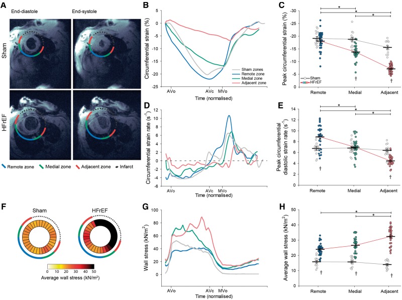
Figure 1.
Slowing of relaxation and elevated wall stress in the adjacent region of HFrEF rats. MRI images illustrate the division of HFrEF rat hearts into three regions (adjacent, medial, and remote) according to their proximity to the infarction (A). Comparison was made to equivalent regions in sham-operated controls. Representative recordings (B) and mean data (C) revealed lower peak circumferential systolic strain adjacent to the infarct in HFrEF. The rate of relaxation was dyssynchronous across the failing heart, as indicated by measurements of circumferential strain rate (D). Specifically, peak diastolic strain rate was only reduced in the adjacent region of HFrEF hearts, and was increased in the remote region (E). Wall stress was calculated across sham and HFrEF hearts based on intraventricular pressure measurements and local ventricular geometry (see Methods section). Measurements are presented as bullseye plots (F) and tracings of wall stress values during the cardiac cycle (G). Mean data demonstrate that while integrated wall stress was high in all regions of HFrEF hearts, values were particularly elevated in the adjacent region (H). (nhearts = 14, 29 in sham, HFrEF). *P < 0.05 calculated by two-way ANOVA with a post hoc Bonferroni t-test. AVc, aortic valve closure; AVo, aortic valve opening; MVo, mitral valve opening.

