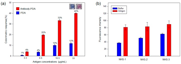
Figure 1.
(a) Colorimetric response (CR%) of the antibody-PDA conjugates and unfunctionalized PDA against antigen concentrations. Inset shows the noticeable color change of antibody-PDA conjugate after the addition of virus antigen (33 µg/mL); (b) Fluorescence intensity of the antibody-PDA conjugates in the absence and presence of virus antigen. (NHS-1 = 10% NHS-PCDA, NHS-2 = 20% NHS-PCDA and NHS-3 = 30% NHS-PCDA) The virus antigen was added to the PDA system and incubated at 37 °C for 10 min.

