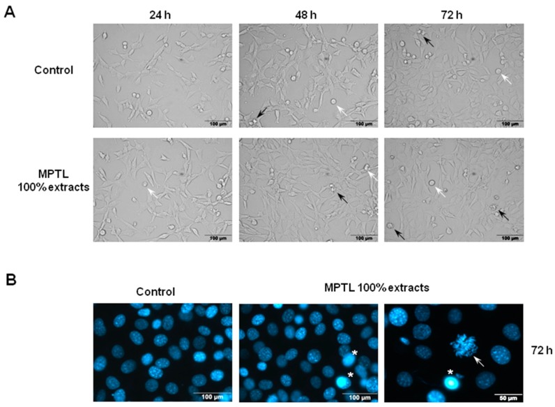
Figure 7.
The effect of MPTL extracts on the: cellular (A); and nuclear (B) morphology of mouse embryonic fibroblast NIH 3T3 cells. (A) NIH 3T3 cells were exposed to 100% concentrated MPTL extract for the time indicated and examined under inverted microscope using ×20 objective. Apart from healthy, flattened adherent cells and detached round mitotic cells (white arrows), there were only few shrunken cells with blebbing membrane typical for apoptotic cell death (black arrows); (B) NIH 3T3 cells were exposed to 100% concentrated MPTL extract for 72 h and examined under fluorescence microscope using ×40 objective following staining with Hoechst 33342. Shrunken nuclei with intensely stained condensed and pycnotic chromatin were regarded as apoptotic (star). Arrow indicates cell with condensed chromosomes undergoing mitosis.

