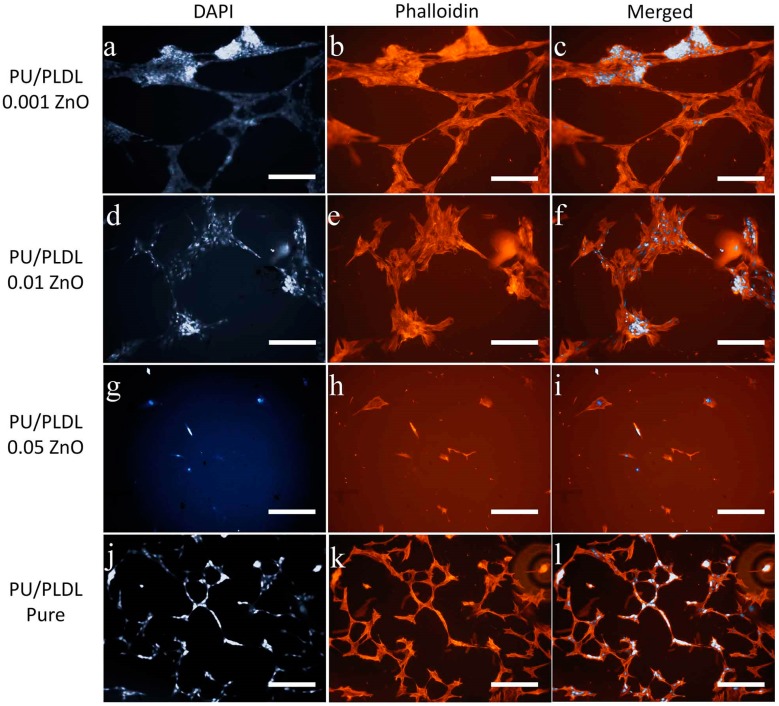
Figure 6.
Adipose stem cells cultured on different experimental polymer substrates stained histochemically for nuclei (DAPI, a,e,i,m), and actin (phalloidin, b,f,j,n), and the cell morphology visualized with SEM (d,h,l,p); cell connections marked with arrowheads; magnifications 100× (fluorescence, scale bar = 200 µm).

