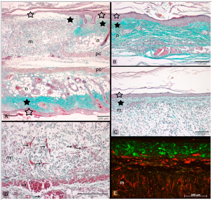
Figure 4.
Skin grafts implanted in mice for 11 days in dorsal skin fold chambers. Sections are stained with Masson’s trichrome (A–D) and analyzed with fluorescence microscopy (E), respectively. (A) Shows the junction between the inserted skin graft (m = Matriderm®) and host mouse skin (n) at the wound edge after 11 days. The engineered and the intact skin part in the sandwiched skin model are separated by the panniculus carnosus (pc). Both in native mouse skin (B) and printed skin graft (C) a thick epidermis (empty asterisks) and corneal layer can be seen. In skin graft, the epidermis layer is formed by the printed keratinocytes (E) noted by green fluorescence that’s emitted by HaCaT-eGFP cells. The fibroblasts (NIH3T3 cells-mCherry) is seen partly migrating into the Matriderm® (yellowish fibres). The fibroblasts that remains on top of the Matriderm®, exhibits an outstretched morphology (C), along with collagen deposition (filled asterisks). Blood vessels (arrows) can be noticed in the skin constructs (D). Scale bars depict 200 µm (A,D,E) and 100 µm (B,C) (Reprinted under open access distribution license from [79]).

