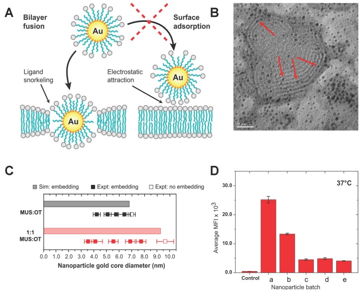
Figure 2.
Penetration of the amphiphilic polymer decorated NPs and its dependence on NP diameter and surface composition. (A) Schematic description of the interaction between amphiphilic polymer-decorated NPs and the cell membrane; (B) Transmission electron microscopy (TEM) image of the giant multilayer vesicle interacting with 1:1 MUS:OT NPs. The red arrows point out that the Au NPs were inserted into the lipid bilayer. The average diameter of the NPs is 2.2 nm; (C) Filled-in squares represent experimental particles that successfully inserted, while empty squares specify those that did not; (D) HeLa cellular uptake in 37 C for all MUS NPs with different diameters, a ( nm), b ( nm), c ( nm), d ( nm), e ( nm). The cellular uptake was measured by the median fluorescence intensity. The figures are adapted from References [52,54] with permission.

