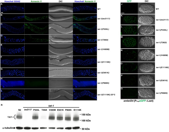
Fig. 3.
PS staining and immunoblotting results of the tat-1 mutants. (A–I) Representative images of dissected gonads from the indicated animals stained with annexin V (green) to visualize externalized PS and Hoechst 33342 (blue) to show nuclei. DIC images of the gonads are also shown. N2 (wild-type) and tat-1(tm3117) strains were included as negative and positive controls, respectively. Images acquired at 20°C (A–H) and 25°C (l) are indicated. (J–Q) Representative images of smIs434 embryos carrying different tat-1 mutations stained for sGFP::Lact (left column) and the corresponding DIC images (right column). Scale bar: 10 μm for all images. (R) Immunoblotting was performed on whole-worm lysates from wild-type or different tat-1 mutant animals as indicated. The mouse monoclonal antibody 7G1 was used to detect TAT-1 proteins (1:1000 dilution). The expression of α-tubulin was used as a loading control. Representative results from at least six independent experiments are shown.

