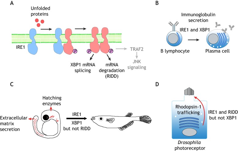
Fig. 1.
The IRE1 branch of the UPR in development and differentiation. (A) A schematic diagram of IRE1 signaling. IRE1 is a transmembrane protein of the ER. Misfolded and/or unfolded proteins in the ER (red circles) promote the oligomerization and trans-autophosphorylation of IRE1, which activate its cytoplasmic RNase function. Active IRE1 splices the mRNA of XBP1 in the cytoplasm to induce stress-responsive gene transcription. In addition, active IRE1 cleaves and degrades a number of other mRNAs through a process that is referred to as RIDD. IRE1 also binds to TRAF2 to activate JNK signaling (gray), but whether this axis plays an active role in animal development remains unclear. (B) IRE1–XBP1 signaling promotes the differentiation of B lymphocytes into plasma cells, which involves expansion of the ER network to allow efficient secretion of immunoglobulins. This differentiation process requires both IRE1 and spliced XBP1. (C) Medaka fish development requires IRE1 and splicing of XBP1 mRNA, but not RIDD. Loss of IRE1 or XBP1 impairs the function of secretory tissues that include the embryonic tail, hatching gland and liver. Spliced XBP1 can rescue the IRE1 mutant phenotype, indicating that the sole function of IRE1 in these tissues is to splice XBP1 mRNA. (D) The requirement for IRE1 in the Drosophila photoreceptor differentiation. Rhodopsin-1 is synthesized in the ER (blue curved bars) and is trafficked to the rhabdomeres, a specialized microvilli-derived structure at the apical membrane (top of the cell). In the absence of IRE1, Rhodopsin-1 fails to traffic properly through the secretory pathway, resulting in photoreceptor differentiation and rhabdomere morphogenesis. This process requires IRE1-mediated RIDD, but not XBP1.

